Unmasking Age Spots: How Senescent Cells Drive Skin Pigmentation
Some links in this article are affiliate links. As an Amazon Associate and partner of other programs, Vitalheros may earn a commission from qualifying purchases, at no extra cost to you. This never influences our editorial coverage.
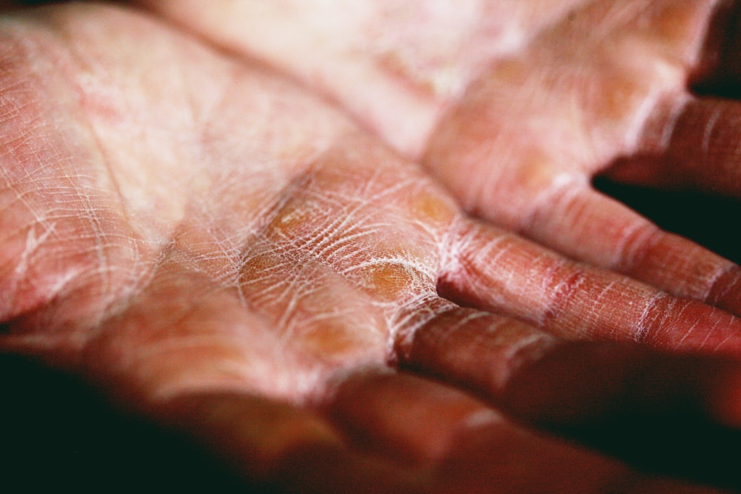
The tell-tale signs of aging often appear first on our skin. Among these, the familiar dark spots commonly known as age spots, or more scientifically, senile lentigo, stand out. These hyperpigmented lesions are a direct consequence of years of sun exposure, a phenomenon known as photoaging. While their appearance is well-known, the underlying cellular mechanisms driving their formation have been a subject of ongoing scientific inquiry.
Recent research is shedding light on a critical player in this process: senescent cells. These ‘zombie cells’ accumulate in tissues over time, contributing to various aspects of aging and age-related diseases. A new study has specifically investigated the presence of these cells within senile lentigo lesions, offering compelling evidence for their role in the development of these common skin changes and pointing towards novel therapeutic avenues.
The Intricacies of Photoaging and Senile Lentigo
Our skin is a remarkable organ, constantly exposed to environmental stressors, with ultraviolet (UV) radiation from the sun being one of the most significant. Chronic UV exposure triggers a cascade of cellular responses that, over time, lead to visible changes such as wrinkles, loss of elasticity, and crucially, hyperpigmentation in the form of age spots.
Senile lentigo presents as distinct, darkened patches on sun-exposed areas like the face, hands, and décolletage. Unlike freckles, which often fade in winter, age spots tend to persist. Their formation involves an increase in melanin production and an altered distribution of melanocytes, the pigment-producing cells in the skin.
The Role of Cellular Senescence in Skin Aging
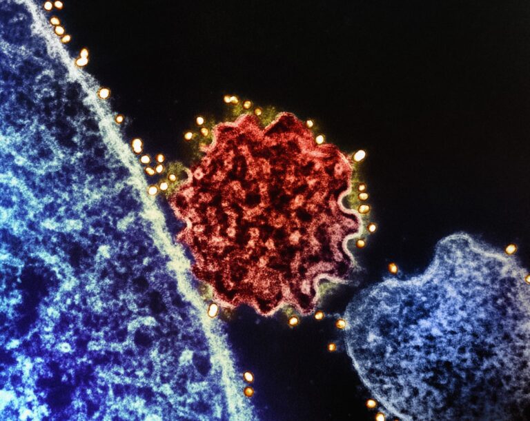
Cellular senescence is a state where cells stop dividing but remain metabolically active, secreting a cocktail of inflammatory molecules, proteases, and growth factors known as the Senescence-Associated Secretory Phenotype (SASP). While initially thought to be a protective mechanism against cancer, the persistent accumulation of senescent cells, particularly in older tissues, is increasingly recognized as a driver of aging and age-related pathology.
In the skin, senescent cells — including fibroblasts, keratinocytes, and melanocytes — have been implicated in various manifestations of aging. Previous studies have linked their accumulation to chronologically aged epidermis, chronic low-dose UV radiation exposure, and even precancerous lesions like actinic keratosis, which are common in sun-damaged skin.
Unveiling Senescent Cells in Age Spots
Recognizing the strong link between UV exposure, photoaging, and senescence, researchers focused their attention on senile lentigo. Given that hyperpigmentation is a prevalent feature of photoaging in certain populations, particularly Asian demographics, the study specifically investigated facial skin biopsy samples from nine Korean donors, classified under Fitzpatrick skin types III or IV.
The researchers collected samples from both lesional sites (the age spots themselves) and adjacent perilesional sites (healthy-looking skin nearby) to provide a direct comparison. As anticipated, the lesional areas exhibited a higher degree of pigmentation and a greater number of melanocytes, confirming the characteristic features of senile lentigo.
Scientific Markers Confirm Senescence
To definitively determine if senescent cells were accumulating in these age spots, the scientists stained the tissue sections for well-established hallmarks of senescence:
- p16INK4A: A cyclin-dependent kinase inhibitor that is a widely used and robust marker of senescence.
- Lamin B1: A nuclear envelope protein whose loss is often associated with cellular senescence.
- Increased nuclear size: A morphological change frequently observed in senescent cells.
Using these specific markers, the study found a significant accumulation of senescent cells within the epidermis of the senile lentigo lesions. This finding is particularly noteworthy because it reinforces earlier observations of senescent keratinocytes in actinic keratosis and chronically UV-exposed skin, suggesting a common underlying mechanism in different forms of sun damage.
Broader Implications and Future Directions
The discovery of increased senescent cell burden in age spots adds a crucial piece to the puzzle of photoaging. It suggests that these ‘zombie cells’ are not merely bystanders but active contributors to the altered structure and function of skin cells within these pigmented lesions. This understanding opens up exciting possibilities for therapeutic interventions.
“The accumulation of senescent cells in senile lentigo underscores their multifaceted role in skin aging, pointing towards senolytics as a promising avenue for improving skin health.”
The Promise of Senolytic Therapies
Senolytic drugs are a class of compounds designed to selectively eliminate senescent cells. In preclinical studies, these agents have shown remarkable potential in reversing various aspects of aging and improving healthspan in animal models. While human data, particularly concerning skin, remains surprisingly limited, there is growing interest in their application to combat photoaging.
Several companies are developing plausibly senolytic skin products, and some senolytic compounds, such as dasatinib and quercetin, are already seeing off-label use. The findings from this study provide a strong rationale for further investigating senolytics as a strategy to not only prevent the formation of age spots but potentially to improve the appearance and health of already aged skin.
Unanswered Questions and Ongoing Research
Despite these advancements, several questions remain. For instance, while the senescence-associated secretory phenotype (SASP) is well-characterized in senescent human fibroblasts, much less is known about the specific SASP profile of senescent keratinocytes, which are abundant in the epidermis. A deeper understanding of these secreted factors could reveal more precise targets for intervention.
Furthermore, the study briefly touches upon Hutchinson-Gilford progeria, a premature aging syndrome linked to altered lamin A and widespread cellular senescence, which also manifests with both hyper- and hypopigmentation. This connection highlights the complex interplay of senescence and pigmentation, suggesting that different cell types within the skin may contribute uniquely to these phenotypes.
Looking Ahead: A New Era for Skin Health
The identification of senescent cells as a key driver of senile lentigo represents a significant step forward in our understanding of skin aging. It reinforces the importance of sun protection as a primary defense against photoaging and lays the groundwork for innovative therapeutic strategies.
As research into senolytics continues to advance, we may soon see targeted treatments that can not only mitigate the cosmetic concerns of age spots but also address the underlying cellular aging processes, leading to genuinely healthier, more resilient skin. The journey from understanding the cellular culprits to developing effective, safe interventions is a complex one, but the potential rewards for longevity and skin health are substantial.
Explore more in our Longevity & Biohacking coverage.
🔬 Scientific Takeaway
New research shows that senile lentigo, commonly known as age spots, contain an increased burden of senescent cells, particularly in the epidermis. This accumulation, linked to chronic UV exposure, suggests that cellular senescence is a key driver of photoaging and the characteristic hyperpigmentation seen in these lesions. The findings bolster the rationale for exploring senolytic therapies as a potential strategy to prevent and even reverse age-related skin changes.
Sources & References
Photo by Alexander Grey on Unsplash.
Medical Disclaimer: This article is AI-assisted and reviewed by the Vitalheros editorial team. It is provided for informational purposes only and is not a substitute for professional medical advice, diagnosis, or treatment. Always consult a qualified healthcare provider. Reviewed by The Vitalheros Editorial Team.