Augmented Reality Transforms Ultrasound Training: Novices Achieve Expert-Level Skill
Some links in this article are affiliate links. As an Amazon Associate and partner of other programs, Vitalheros may earn a commission from qualifying purchases, at no extra cost to you. This never influences our editorial coverage.
In the demanding world of medical diagnostics, expertise is often honed over years of rigorous training and hands-on experience. Mastering complex imaging techniques, such as ultrasound, requires not only deep anatomical knowledge but also a highly developed spatial awareness and psychomotor skill. However, a groundbreaking convergence of real-time 3D ultrasound and augmented reality (AR) is now dramatically shortening this learning curve, empowering even novice practitioners to perform with the precision typically associated with seasoned experts.
This innovative approach represents a significant leap forward in medical education and clinical practice. By seamlessly blending digital information with the physical world, AR is poised to democratize access to high-quality diagnostic imaging, ensuring that critical skills are acquired more rapidly and effectively than ever before.
The Challenge of Ultrasound Mastery
Ultrasound imaging is a cornerstone of modern medicine, providing non-invasive, radiation-free insights into the body’s internal structures. From monitoring fetal development to diagnosing cardiovascular conditions, its versatility is unmatched. Yet, its operator-dependent nature presents a steep learning curve. Unlike X-rays or CT scans, which produce static images, ultrasound requires the practitioner to actively manipulate a probe to generate and interpret dynamic, two-dimensional slices of a three-dimensional anatomy.
- Spatial Reasoning: Translating 2D images into a mental 3D model.
- Probe Manipulation: Achieving the correct angle, pressure, and movement for optimal imaging.
- Image Interpretation: Recognizing subtle pathological changes amidst normal anatomy.
- Real-time Adaptation: Adjusting scanning techniques based on live feedback.
Traditionally, acquiring these skills demands extensive supervised practice, often involving long hours in a clinical setting under the watchful eye of an experienced sonographer. This resource-intensive training model can limit the number of practitioners who achieve proficiency, particularly in areas with fewer expert trainers.
Augmented Reality: A New Dimension in Medical Training
Augmented reality technology overlays digital content onto a user’s view of the real world, enhancing perception and interaction. Think of it as a sophisticated digital layer that provides context-sensitive information exactly when and where it’s needed. When combined with real-time 3D ultrasound data, AR transforms the learning process into an intuitive, guided experience.
How it Works: Seeing Through the Skin
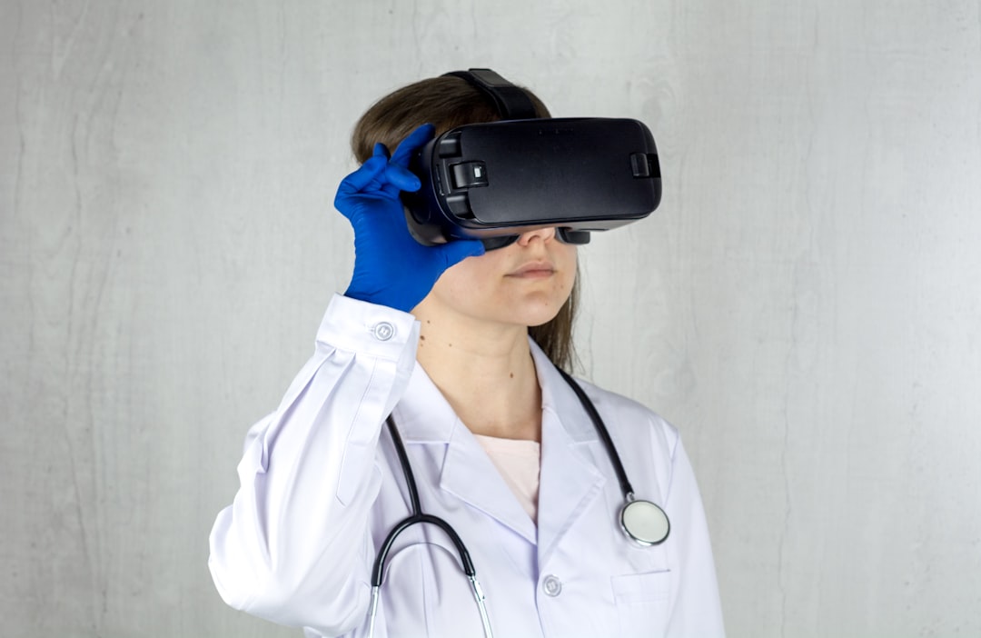
Imagine a medical trainee wearing an AR headset. As they hold an ultrasound probe, the headset projects a dynamic, three-dimensional reconstruction of the patient’s internal organs directly onto the patient’s body or into the trainee’s field of vision. This isn’t just a static image; it’s a real-time, interactive guide.
The system can:
- Visualize Internal Anatomy: Display the target organ (e.g., heart, kidney, blood vessel) as a transparent overlay on the patient’s skin, showing its exact position and orientation.
- Guide Probe Placement: Provide visual cues and arrows indicating the optimal angle and position for the ultrasound probe to capture the desired view.
- Highlight Key Structures: Emphasize important anatomical landmarks or areas of interest, helping the trainee focus their scan.
- Offer Real-time Feedback: Instantly assess the quality of the scan and provide corrective guidance, much like an expert instructor would.
- Display 3D Ultrasound Data: Integrate the live 2D ultrasound images into a comprehensive 3D model, allowing trainees to better understand the relationship between their probe movements and the resulting imagery.
This innovative setup effectively allows the trainee to
Explore more in our Digital Health coverage.
🔬 Scientific Takeaway
The integration of real-time 3D ultrasound with augmented reality (AR) significantly enhances medical training, particularly for complex diagnostic procedures like ultrasound imaging. By overlaying dynamic anatomical models and guidance directly into a user's field of vision, AR technology enables novice practitioners to quickly develop the spatial reasoning and psychomotor skills necessary to perform at a level comparable to experienced experts. This innovation promises to accelerate skill acquisition, potentially broadening access to high-quality diagnostic capabilities across various healthcare settings.
Sources & References
Photo by Bermix Studio on Unsplash.
Medical Disclaimer: This article is AI-assisted and reviewed by the Vitalheros editorial team. It is provided for informational purposes only and is not a substitute for professional medical advice, diagnosis, or treatment. Always consult a qualified healthcare provider. Reviewed by The Vitalheros Editorial Team.