Engineered Scaffold Offers New Hope for Craniosynostosis Treatment
Some links in this article are affiliate links. As an Amazon Associate and partner of other programs, Vitalheros may earn a commission from qualifying purchases, at no extra cost to you. This never influences our editorial coverage.
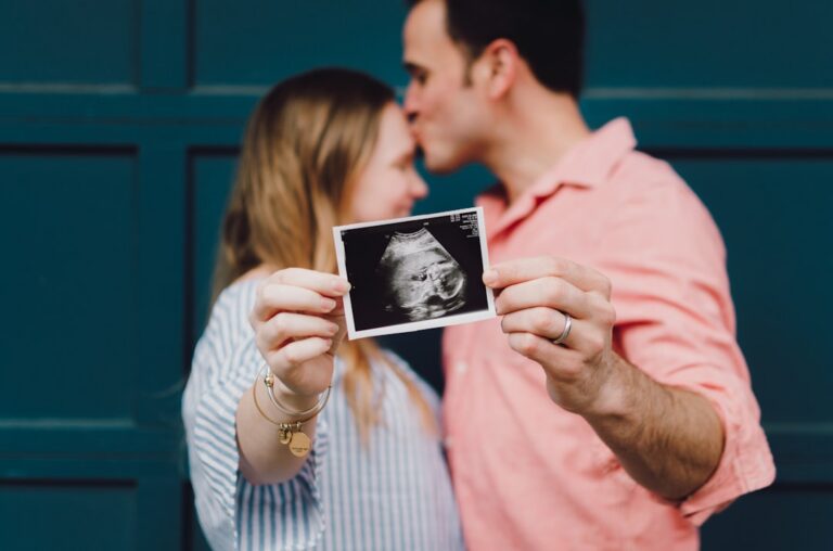
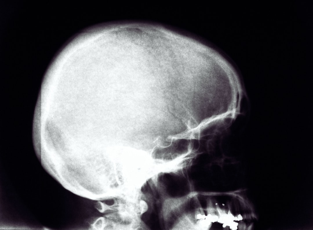
Craniosynostosis, a condition where a baby’s skull bones fuse prematurely, presents significant challenges for affected children and their families. This early fusion can restrict brain growth, leading to developmental issues, increased intracranial pressure, and facial deformities. While surgical intervention is the standard of care, it often carries risks and, in some cases, may not fully prevent the need for further procedures. Now, groundbreaking research involving an engineered scaffold is offering a beacon of hope, demonstrating the potential to restore normal skull growth in craniosynostosis mouse models.
Understanding Craniosynostosis: A Complex Challenge
Normally, a baby’s skull is composed of several separate plates of bone, connected by flexible sutures. These sutures allow the skull to expand as the brain grows rapidly during infancy and early childhood. In craniosynostosis, one or more of these sutures close too soon, before the brain has finished growing. This premature fusion dictates the shape of the skull, often leading to an abnormal head shape as the brain tries to grow in the path of least resistance. Beyond cosmetic concerns, the restricted growth can place pressure on the developing brain, potentially causing:
- Developmental delays
- Learning difficulties
- Vision problems
- Headaches
- Seizures
Current Treatment Limitations
The primary treatment for craniosynostosis is surgery, which aims to separate the fused bones and reshape the skull to allow for proper brain growth. While often effective, these complex procedures are typically performed in infancy and carry inherent risks, including blood loss and infection. Furthermore, in some cases, the bones may re-fuse or the skull may not achieve optimal shape, necessitating additional surgeries. The quest for less invasive, more effective, and longer-lasting solutions has long been a priority for pediatric neurosurgeons and researchers.
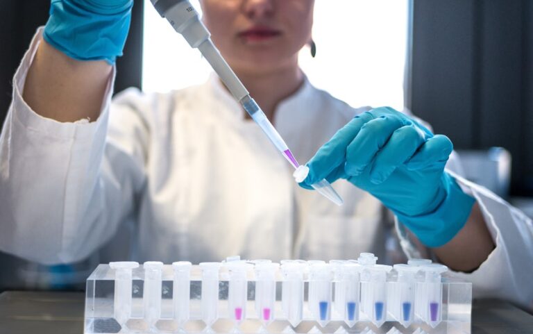
A Novel Approach: Engineered Scaffolds for Bone Regeneration
The new research introduces an innovative strategy that moves beyond simply separating fused bones. Instead, it focuses on guiding the skull’s natural regenerative processes using a specially designed engineered scaffold. This approach harnesses principles of regenerative medicine, aiming to create an environment where the skull can effectively ‘relearn’ how to grow correctly.
How the Scaffold Works
At its core, the engineered scaffold is a sophisticated biological and mechanical construct. While specific details of its composition would depend on the research, such scaffolds typically involve:
- Biocompatible Materials: The scaffold is made from materials that are safe for the body and can either degrade harmlessly over time or integrate into existing tissue.
- Porous Structure: Its intricate, porous design mimics the natural extracellular matrix, providing a framework for new bone cells to migrate into, attach, and proliferate.
- Growth Factor Delivery: Often, these scaffolds are loaded with specific growth factors or signaling molecules. These biological cues act like instructions, guiding stem cells and other progenitor cells to differentiate into bone-forming cells (osteoblasts) and lay down new, healthy bone tissue.
- Mechanical Support: The scaffold provides temporary structural support, helping to maintain the desired skull shape while the new bone grows, preventing premature re-fusion.
The idea is not just to fill a gap, but to actively orchestrate the biological processes that lead to controlled, directed bone formation, essentially teaching the skull how to grow in a healthy pattern.
Promising Results in Craniosynostosis Mouse Models
The most compelling aspect of this research lies in its outcomes within craniosynostosis mouse models. These models are crucial for understanding the disease and testing new therapies before human trials. In these studies, the engineered scaffold demonstrated a remarkable ability to restore normal skull growth patterns.
The findings indicate that the scaffold successfully facilitated the regeneration of functional sutures, allowing the skull to expand symmetrically and accommodate brain growth, much like a healthy skull would. This suggests the scaffold could overcome the critical challenge of re-fusion that often plagues traditional surgical approaches.
By preventing premature fusion and guiding appropriate bone formation, the scaffold effectively normalized the cranial development in the animal models. This is a significant step forward, as it tackles the root problem of restricted growth rather than merely correcting its consequences.
Implications for Future Pediatric Care
While these results are from mouse models and extensive further research is needed, the implications for human craniosynostosis treatment are profound:
- Reduced Re-operation Rates: If successful in humans, this technology could significantly decrease the need for repeat surgeries, easing the burden on children and their families.
- Improved Developmental Outcomes: By ensuring more natural and unrestricted brain growth, the scaffold could potentially lead to better cognitive and neurological development.
- Less Invasive Options: While still a surgical intervention, the scaffold-based approach might offer a more biologically integrated solution compared to current methods, potentially leading to better long-term outcomes.
- Personalized Medicine: In the future, it might be possible to engineer scaffolds tailored to the specific needs of each child, optimizing bone regeneration based on their unique skull anatomy and fusion pattern.
Looking Ahead: From Bench to Bedside
It is important to emphasize that research in mouse models represents an early, albeit critical, stage of development. Translating these findings to human clinical application will require rigorous testing, including preclinical studies in larger animal models, followed by carefully designed human clinical trials. This process can take many years.
However, the promise of an engineered scaffold to fundamentally restore normal skull growth in craniosynostosis is an exciting prospect. It underscores the power of regenerative medicine and biomedical engineering to address complex congenital conditions, offering a glimmer of hope for a future where children born with craniosynostosis can achieve healthier, more typical development with fewer interventions.
Explore more in our Digital Health coverage.
🔬 Scientific Takeaway
An engineered scaffold has shown promising results in restoring normal skull growth in craniosynostosis mouse models. By providing a framework for bone regeneration and delivering growth-promoting signals, the scaffold facilitated the formation of functional sutures, allowing for symmetrical skull expansion. This innovative approach holds potential to overcome limitations of current surgical interventions and improve developmental outcomes for children with craniosynostosis.
Sources & References
Photo by Risto Kokkonen on Unsplash.
Medical Disclaimer: This article is AI-assisted and reviewed by the Vitalheros editorial team. It is provided for informational purposes only and is not a substitute for professional medical advice, diagnosis, or treatment. Always consult a qualified healthcare provider. Reviewed by The Vitalheros Editorial Team.