How Your Visual Environment Reshapes the Eye’s Retina
Some links in this article are affiliate links. As an Amazon Associate and partner of other programs, Vitalheros may earn a commission from qualifying purchases, at no extra cost to you. This never influences our editorial coverage.
Our eyes are often described as windows to the world, but new research suggests the world itself might be subtly reshaping our eyes. A recent study, leveraging the transparent nature and well-understood visual system of zebrafish, has shed light on a fascinating aspect of ocular biology: the profound plasticity of the retina. This research indicates that the visual environment we inhabit can directly influence the structural organization of this critical part of the eye, offering new insights into how vision develops and adapts.
For decades, scientists have understood that our brains exhibit remarkable plasticity, constantly reorganizing neural pathways in response to experience. However, the extent to which the eye itself, particularly the retina – the light-sensing tissue at the back of the eye – can undergo structural adaptation based on visual input has been a subject of ongoing inquiry. This latest work provides compelling evidence that the retina is far from a static structure; it actively responds to its visual surroundings, fundamentally altering its architecture.
The Retina: A Dynamic Canvas of Vision
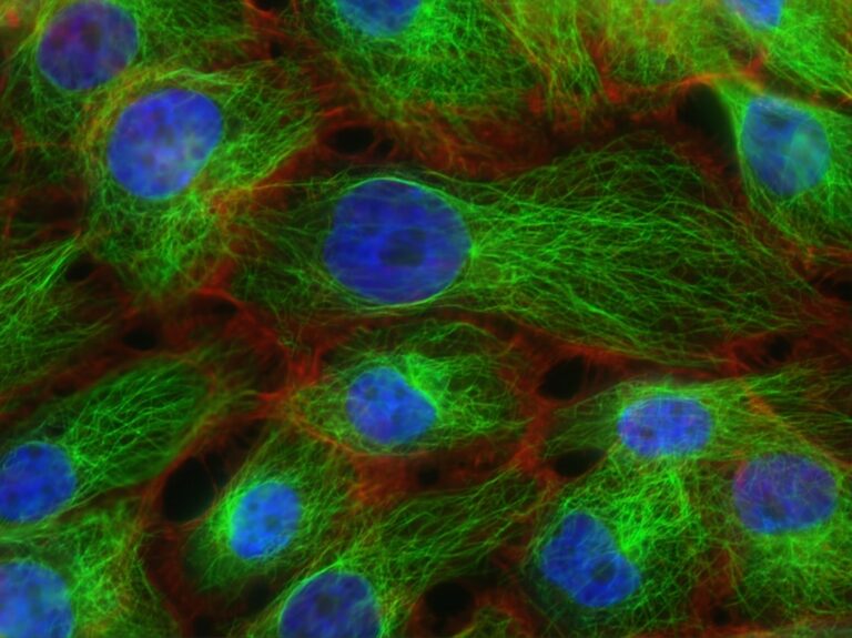
The retina is an intricate network of specialized cells, including photoreceptors that detect light and various types of neurons that process visual information before sending it to the brain. It’s often considered the ‘film’ of the camera-like eye, capturing images and initiating the complex process of sight. While its basic structure is genetically determined, mounting evidence suggests it’s also a highly adaptable tissue, capable of fine-tuning its organization in response to external cues.
This adaptability is crucial for optimal vision. Imagine a world where our eyes couldn’t adjust to varying light conditions or different visual demands. Our ability to perceive depth, focus on objects at different distances, and distinguish colors relies on a retina that can dynamically respond to its environment. The new research delves into the very structural underpinnings of this adaptability.
Why Zebrafish Are an Ideal Model
To unravel the intricate mechanisms behind retinal reshaping, researchers often turn to model organisms. Zebrafish (Danio rerio) have emerged as particularly valuable for studying vision and ocular development for several key reasons:
- Transparency: Young zebrafish embryos and larvae are largely transparent, allowing scientists to observe internal structures, including the developing eye and retina, in real-time without invasive procedures.
- Genetic tractability: Their genetics are well-understood, and researchers can easily manipulate genes to study their roles in development and disease.
- Rapid development: Zebrafish develop quickly, enabling studies on developmental processes over relatively short periods.
- Retinal similarity: Despite being fish, their retinas share significant structural and functional similarities with mammalian retinas, including the presence of rods and cones for light detection and a complex neuronal circuitry for processing.
These characteristics make zebrafish an excellent living laboratory for observing how environmental factors, specifically visual input, can mold the eye’s architecture.
How Visual Cues Influence Retinal Structure
The study demonstrated that simply altering the visual environment of zebrafish could induce measurable structural changes within their retinas. While the precise details of these changes (e.g., specific cell types or neuronal connections) were not fully elaborated in the initial summary, the core finding points to a fundamental principle: the visual input received by the eye is not merely passively processed; it actively participates in shaping the very sensory organ that receives it.
This suggests a feedback loop where the information perceived by the eye influences its physical makeup, potentially optimizing it for the specific visual challenges of its surroundings. For instance, an environment rich in fine details might encourage the development of retinal areas with higher acuity, while an environment dominated by motion might enhance areas responsible for detecting movement.
Beyond Simple Adaptation: Implications for Visual Processing
These structural alterations are not just cosmetic; they likely have profound implications for how visual information is processed. A reshaped retina could mean altered neuronal pathways, different densities of specific cell types, or modifications in the strength of synaptic connections. Such changes could lead to a retina that is, in essence, ‘tuned’ to its prevalent visual input, enhancing its efficiency and accuracy in that specific environment.
This dynamic interplay between environment and structure highlights the eye as a remarkably plastic organ, capable of self-optimization throughout development and, perhaps, even into adulthood. Understanding these mechanisms could provide a deeper insight into the fundamental principles governing sensory system development.
Potential Implications for Human Vision and Health
While this research was conducted in zebrafish, its findings open intriguing avenues for understanding human eye development and conditions. The concept that visual surroundings can structurally reshape the retina carries significant implications, particularly for refractive errors like myopia (nearsightedness) and hyperopia (farsightedness).
Myopia, for example, is reaching epidemic proportions globally, and environmental factors, such as increased near work and reduced time outdoors, are strongly implicated in its development. While the primary mechanism for myopia is often linked to the lengthening of the eyeball, the retina’s role in sensing and responding to visual cues is critical. If different visual environments can structurally alter the retina, it’s plausible that these changes could contribute to, or be a consequence of, the eye’s axial growth and refractive state.
This foundational research underscores the sophisticated ways our sensory organs interact with the world, offering a glimpse into how targeted environmental interventions or therapies might one day be developed to support healthy vision.
<
Future Directions and Therapeutic Potential
This zebrafish study serves as a critical piece of foundational research. The next steps would involve identifying the specific molecular and cellular pathways involved in this retinal reshaping. Pinpointing these mechanisms could unlock potential therapeutic targets for preventing or managing vision disorders that have an environmental component.
For instance, if certain light patterns or visual stimuli are found to promote healthy retinal development or mitigate adverse structural changes, this knowledge could inform strategies for early childhood visual environments, educational practices, or even novel light therapies. It reinforces the idea that our visual health is not solely determined by genetics but is an ongoing dialogue between our inherent biology and the world we perceive.
The intricate dance between our eyes and our environment is far more complex and dynamic than previously appreciated. This research in zebrafish reminds us that the quest to understand vision is a journey into the remarkable adaptability of life itself.
Explore more in our Digital Health coverage.
🔬 Scientific Takeaway
A zebrafish study reveals the retina is a highly plastic organ, with its structural organization being actively reshaped by the visual environment. This suggests a dynamic feedback loop where visual input influences the physical architecture of the eye. The findings offer foundational insights into sensory system development and may have future implications for understanding human vision disorders influenced by environmental factors.
Sources & References
Photo by Wolfgang Hasselmann on Unsplash.
Medical Disclaimer: This article is AI-assisted and reviewed by the Vitalheros editorial team. It is provided for informational purposes only and is not a substitute for professional medical advice, diagnosis, or treatment. Always consult a qualified healthcare provider. Reviewed by The Vitalheros Editorial Team.